Spatiotemporal mapping of the contractile and adhesive forces sculpting early C. elegans embryos - Developmental Cell
K. Yamamoto, S. Ichbiah, M. Perez, J. Borrego-Pinto, F. Delbary, N. Goehring, P. Northrop, H. Turlier*, G. Charras*. Developmental Cell, 61, 1-16 (2026)
Read more: publisher - preprint
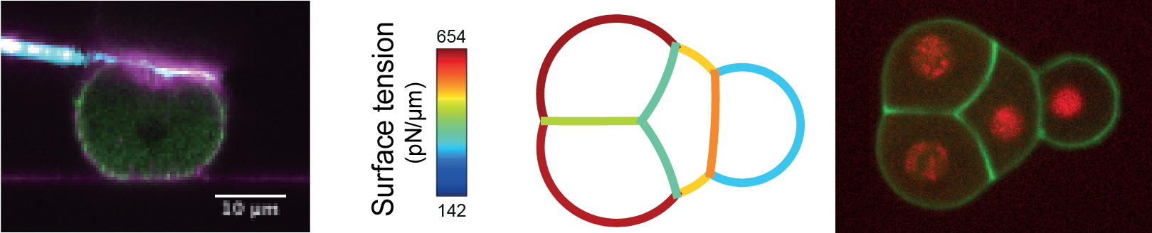
Embryo shape is determined by cell mechanics, intercellular interactions, and geometrical constraints. Models based on surface tensions at cell interfaces can predict 3D static cellular arrangements within aggregates. However, predicting the dynamics of such arrangements is challenging because temporal changes in tension are unknown. Here, we characterize the spatiotemporal changes in cellular tensions shaping early nematode embryos using atomic force microscopy (AFM), live microscopy, and tension inference. Using excoriated embryos, we validate a hybrid inference pipeline that temporally calibrates relative inferred tensions using cortical myosin enrichment and absolute tensions obtained by AFM. Applying this approach to embryos within their native shell provides a spatiotemporal map of absolute tensions. This reveals that ABa, ABp, and EMS compaction is driven by increased cell-medium tension, whereas P2’s initial exclusion is due to high cell-cell contact tension. We further uncover a direct contribution of cadherins to cell-cell contact tension, comparable in magnitude to cadherins’ indirect contribution via actomyosin regulation.